APRT Deficiency / 2,8-DHA crystalluria
High resolution photomicrographs of 2,8-DHA Crystals
Click on an image to see the full resolution image (1600 x 1200 px). Images show 2,8-Dihydroxyadenine crystals in the urine sediment. Conventional light (brightfield) microscopy is used unless noted otherwise.
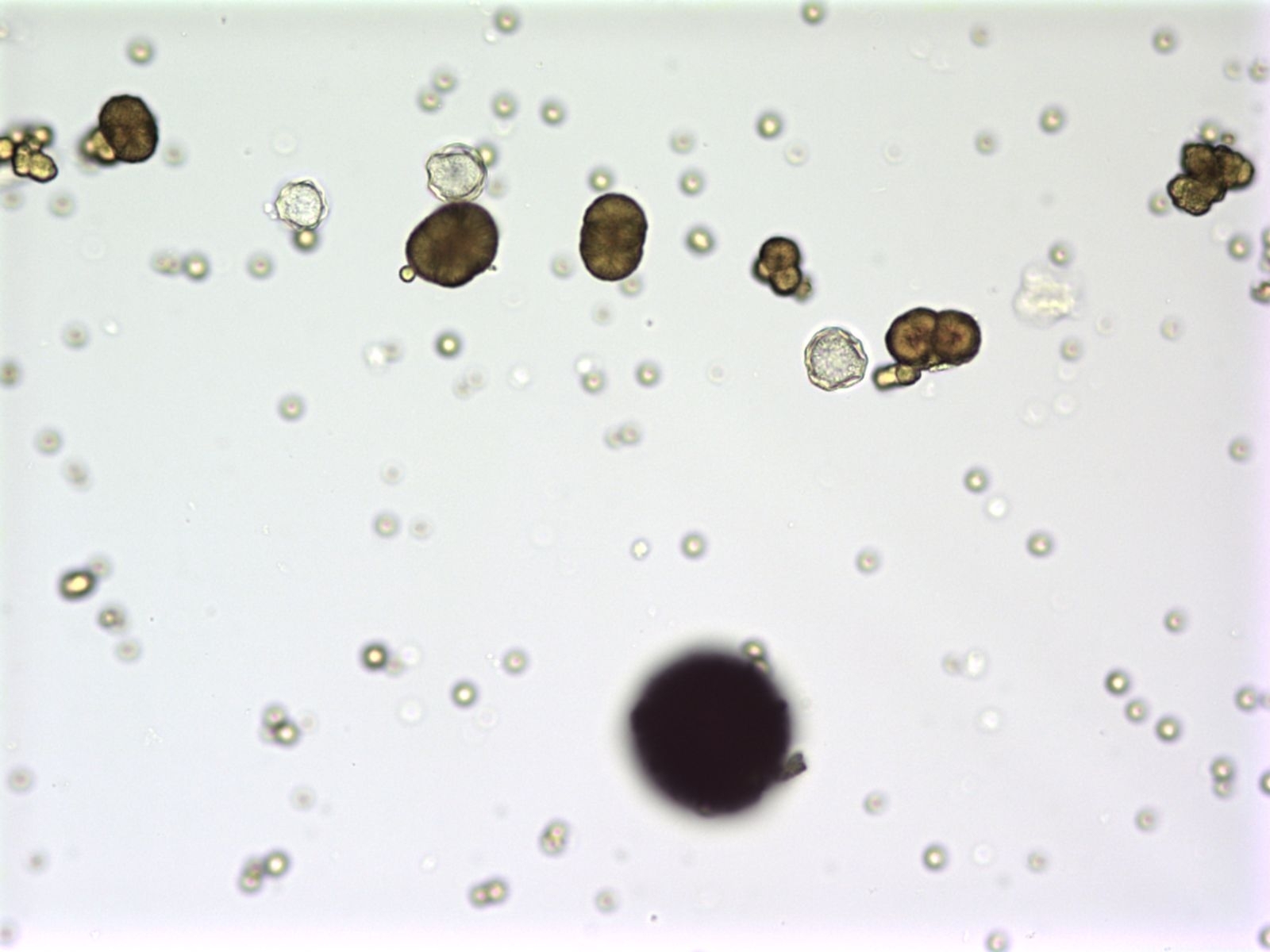
Figure 1a. Several medium sized 2,8-DHA crystals and many small sized crystals are present. One large crystal at the bottom of the figure is out of focus. (Original magnification x 400).
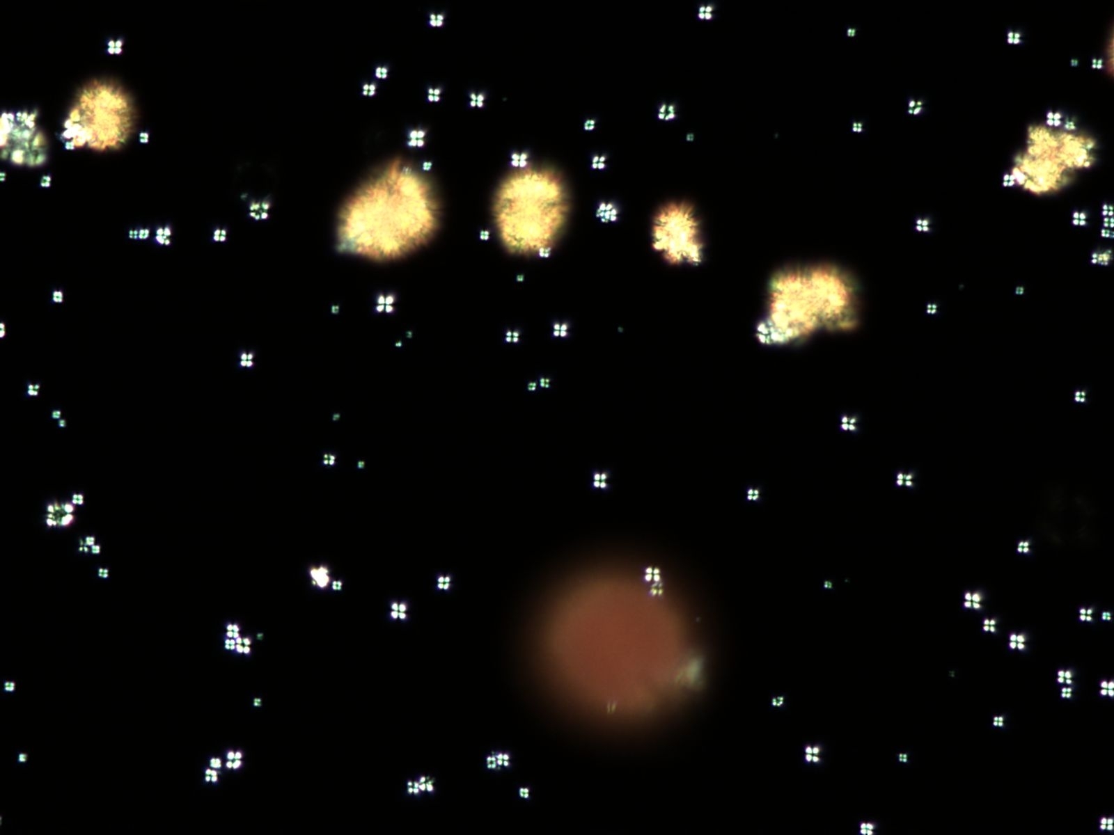
Figure 1b. The same field as in Figure 1a viewed by polarized light microscopy. The small sized 2,8-DHA crystals appear white and produce the characteristic Maltese cross pattern. The medium sized crystals appear yellow. (Original magnification x 400).
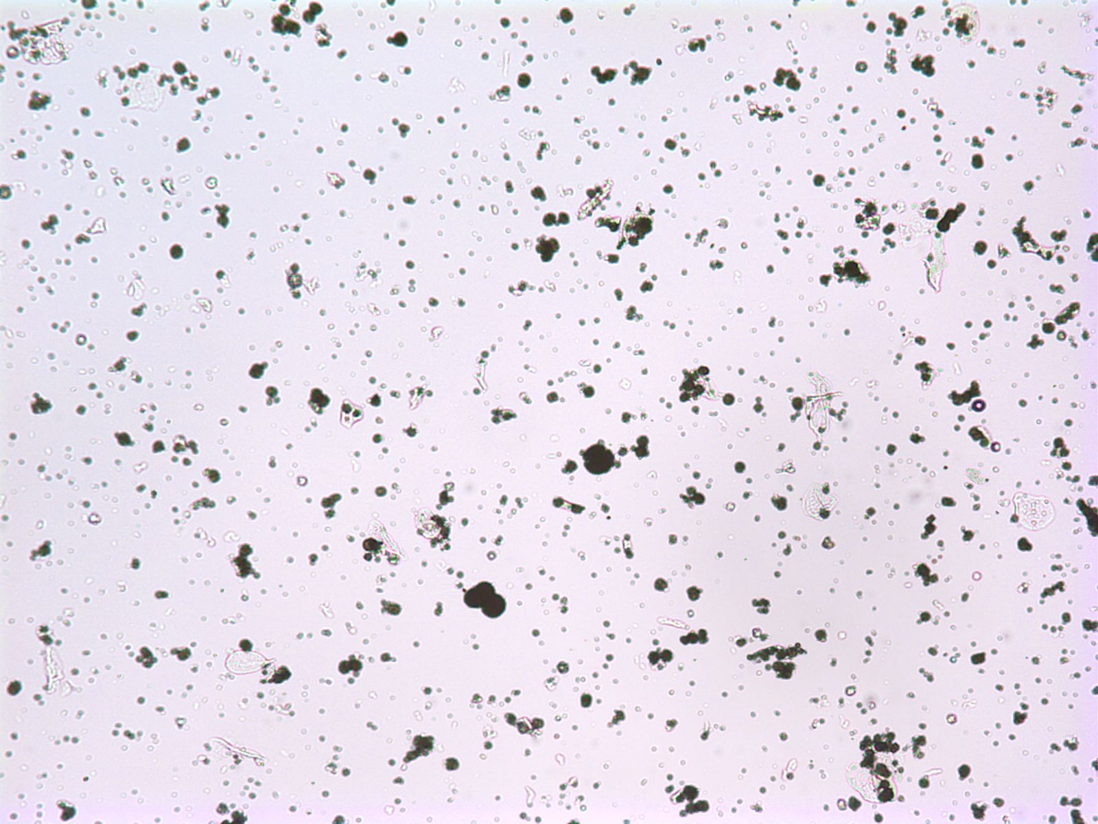
Figure 2. Different sizes of 2,8-DHA crystals. The larger crystals appear dark. (Original magnification x 100).
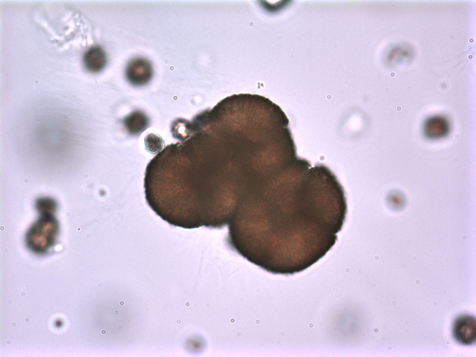
Figure 3. Typical round, reddish-brown 2,8-DHA crystals. Note the dark outline, central density and the radiating spicules. (Original magnification x 1000).
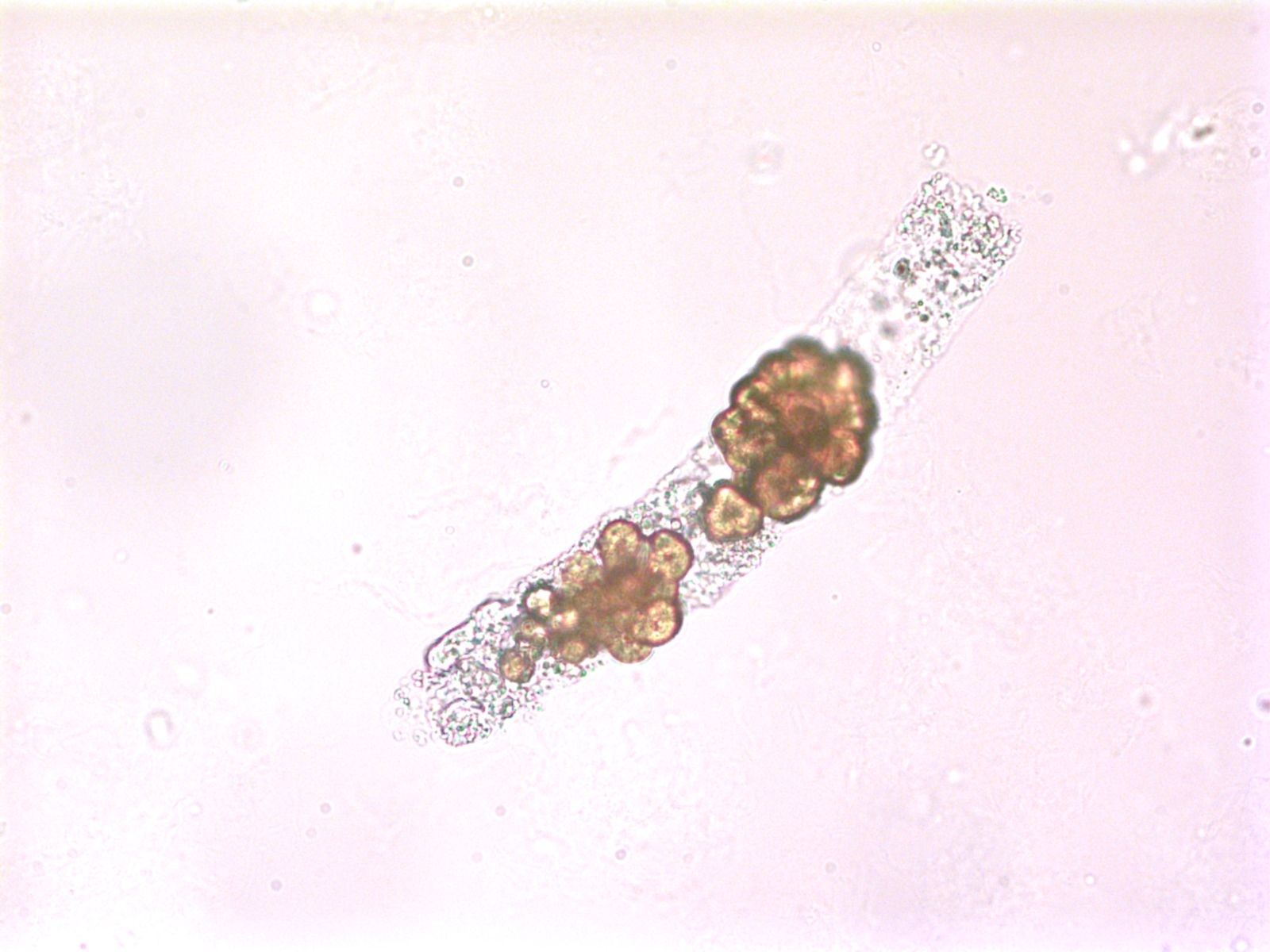
Figure 4. 2,8-DHA crystals are seen within a granular cast. (Original magnification x 400).
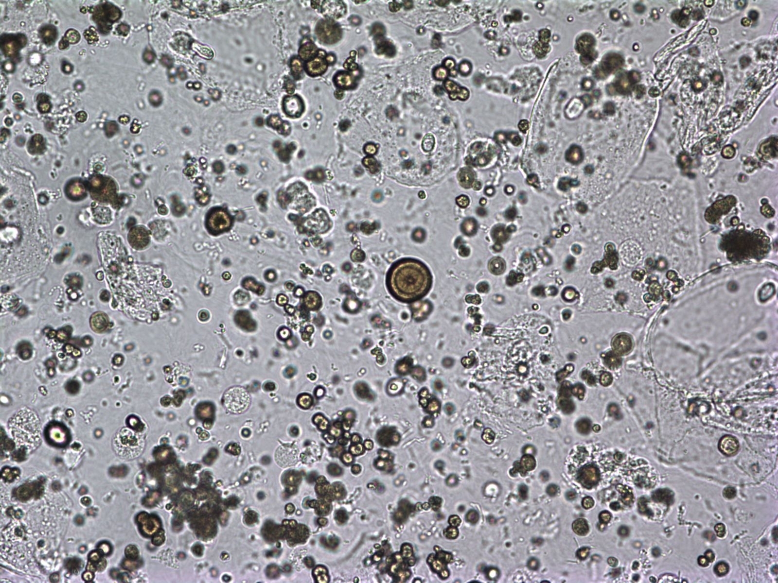
Figure 5. 2,8-DHA crystals of variable sizes. In the middle of figure there is a crystal showing a tree-ring pattern. (Original magnification x 400).
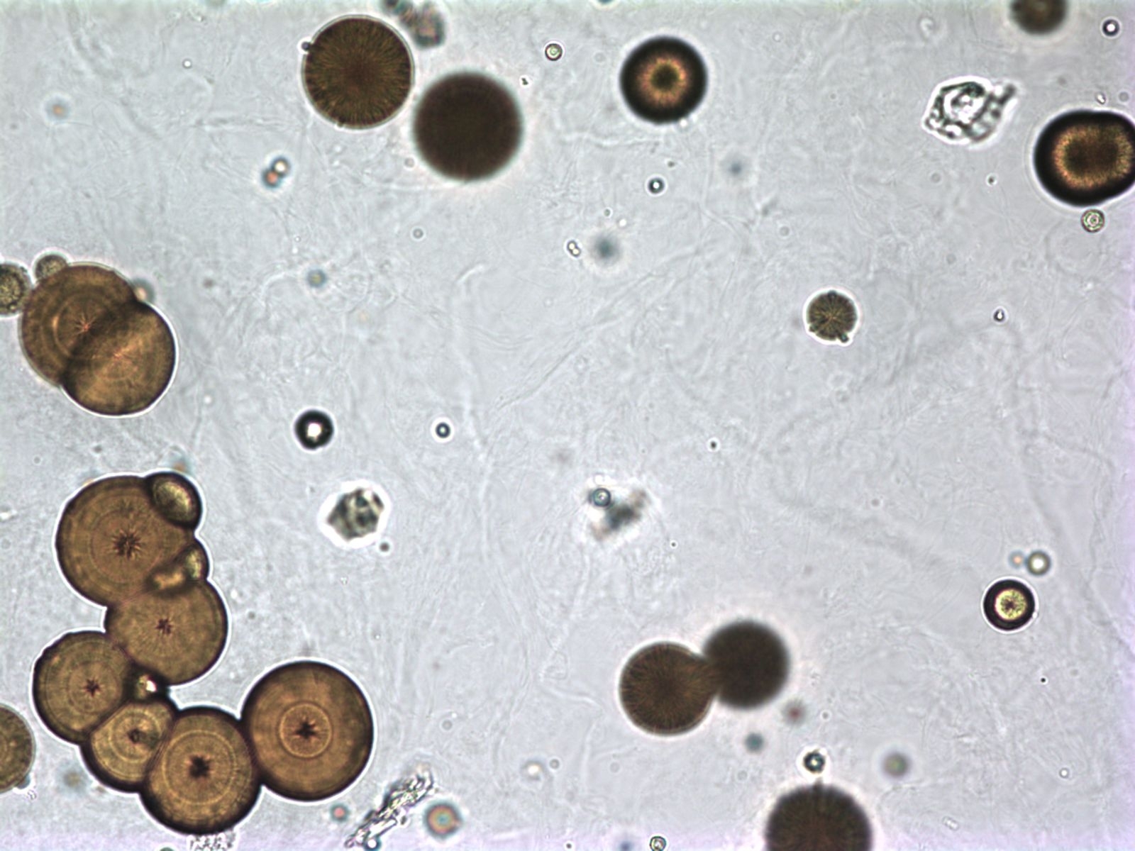
Figure 6. Medium-sized 2,8-DHA crystals. Note the tree-ring pattern of the crystals on the left side of the figure. (Original magnification x 400).
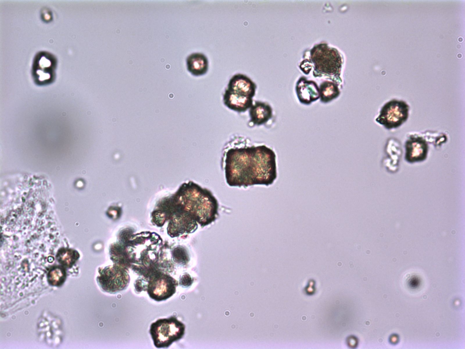
Figure 7. 2,8-DHA crystals demonstrating a rarely seen rectangular or square shape. (Original magnification x 1000).
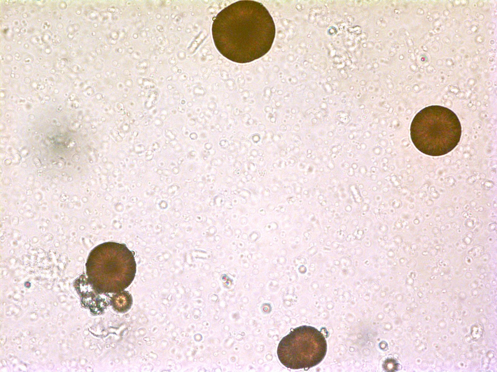
Figure 8. Four typical, round and reddish-brown 2,8-DHA crystals. Note the central density, radiating central spicules and the dark outline. (Original magnification x 400).

Figure 9. Several 2,8-DHA crystals with extra ordinary clear ring-tree pattern. The largest crystal is characteristically reddish in colour. Calcium oxalate and uric acid crystals are also present. (Original magnification x 400).
Acknowledgements
Photomicrographs of 2,8-dihydroxyadenine crystals were kindly provided by Gudridur Steinunn Oddsdottir, Biomedical Scientist, Departments of Laboratory Hematology and Clinical Biochemistry, Landspitali University Hospital, Reykjavik, Iceland.
 Consortium Home
 APRT Deficiency